Creating a Masterpiece, One Ligament at a Time
As Modernizing Medicine’s Medical Illustrator, Taryn Baacke holds one of the company’s more unique roles with her influence on our Electronic Medical Assistant® (EMA ). She creates the EMA 3D Interactive Anatomical Atlas, a zoomable, high-resolution anatomic body illustration to assign diagnoses and treatments and characterize findings. Taryn sat down for an interview to share what it takes to develop this multi-view, three-dimensional layered tool.
Q: As the Medical Illustrator, what exactly do you do?
Taryn: I get asked that question a lot! The answer is that it depends on which specialty-specific EMR system I’m working on and the day. I use Adobe Photoshop to create new images, i.e. body zoning for cutaneous innervation. I then move that image into Adobe Illustrator to develop the images you actually see in EMA’s Virtual Exam Room.
Q: What was your career path? Which came first: your eye for medicine or your eye for artistic beauty?
Taryn: Neither! My original bachelor’s degrees were in marine biology and art. I always wanted to combine science and art – I love both disciplines and didn’t want to have to decide between the two. A college advisor asked me to create an image for an invertebrate anatomy class, and then suggested that I do this for a living after attending graduate school. Classes I studied during my Master’s of Fine Arts degree included medical illustration, surgical observation, cadaver dissection for gross anatomy, art classes, Photoshop and Illustrator, to name a few.
Q: Why should doctors care about the EMA 3D Interactive Anatomical Atlas and how it’s created?
Taryn: The EMA 3D Interactive Anatomical Atlas is geared towards a specific patient – male or female, adult or pediatric. The images are as medically accurate as possible for each specialty because we have physicians on staff. For example, I consult with two of our orthopedic surgeons, Matt Stiebel and Dan Prince, so the skeleton view in EMA Orthopedics isn’t simply broken up into bones; it’s broken into fracture patterns based on how each individual bone breaks most often.
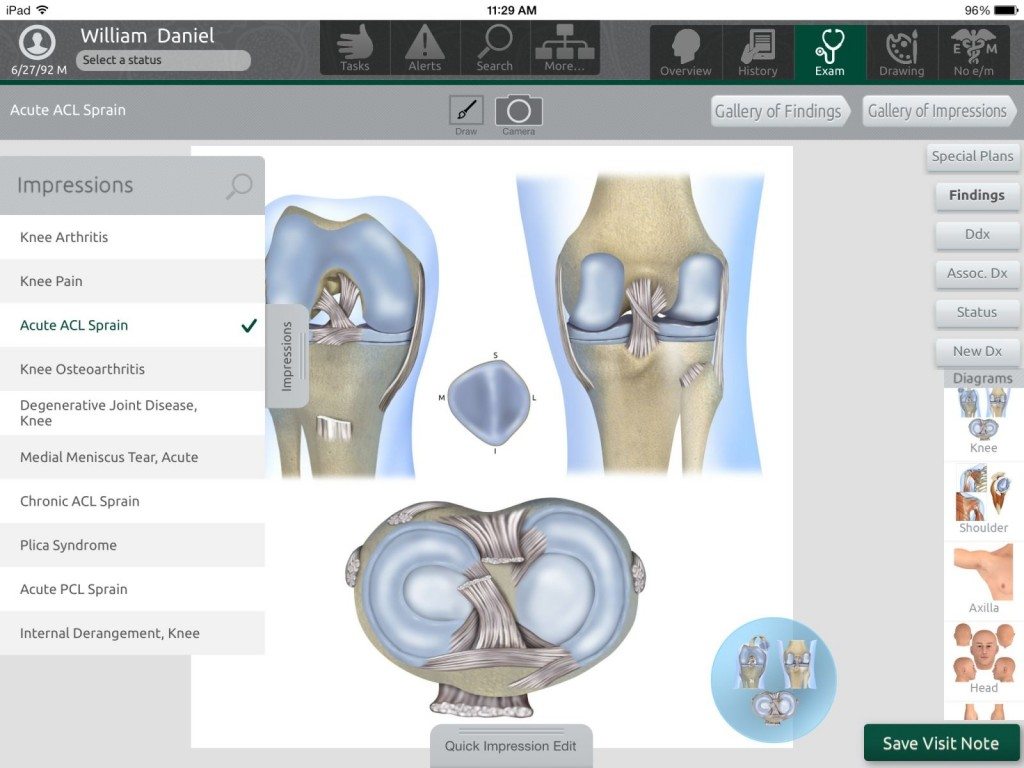
The EMA 3D Interactive Anatomical Atlas includes a 360 view that details even the top of the head and bottom of the feet. Users can zoom in to see very detailed drawings of ligaments and soft tissue of the knee
The EMA 3D Interactive Anatomical Atlas includes a 360 view that details even the top of the head and bottom of the feet. Users can zoom in to see very detailed drawings of ligaments and soft tissue of the knee (see below).
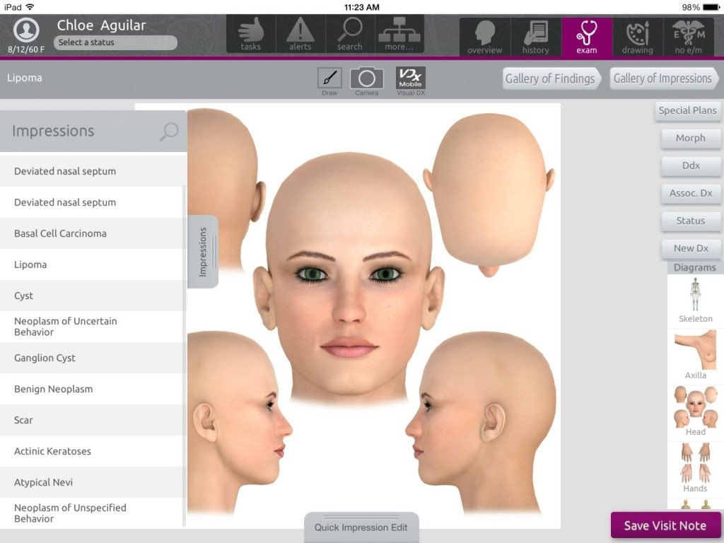
Dermatologists using EMA Dermatology can see in one image a compilation of all angles of the face: front, back, left, right and top of head (see below).
Dermatologists using EMA Dermatology can see in one image a compilation of all angles of the face: front, back, left, right and top of head
Our clients requested this feature as well as images of the front and back of hands and the nail beds. We appreciate user feedback and incorporate it whenever possible.
The level of detail of the data captured helps make exam notes more specific. Physicians using such advanced technology don’t have to copy and paste, which is not recommended. Because it’s so easy to use, EMA is a time saver enabling physicians to spend more time with patients and less time documenting.
Q: What is your personal goal?
Taryn: I would love to see EMA evolve into more of a tool for physicians to educate patients, as EMA is already used educationally. One fantastic example is the case when an ophthalmologist using EMA Ophthalmology contacted us and wanted specific views of the eye in the eye socket. He wanted to draw on the images in the Drawing Room to show patients what he was doing and where he was doing it. These images can then be attached to the patient’s chart in EMA and assigned to that specific visit. Our ophthalmology clients love this feature.