5 Must-Have Ophthalmology Image Management Software Features
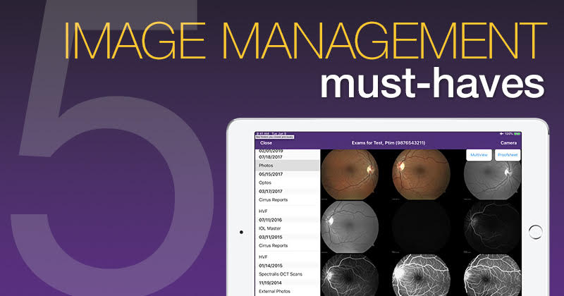
Here’s what can set ophthalmic image management software apart from the crowd
Ophthalmology is an extremely visual field (no pun intended). Much of the work you do revolves around looking at images from diagnostic devices. But it shouldn’t take sifting through stacks of papers or clicking 30 times to pull up the image you need.
This is where ophthalmology software comes in. A good ophthalmic image management system will help make it easy for you to import, find, view and manipulate images. But watch out for inferior systems that can slow you down instead of helping you.
Here are my top five features that help set the best ophthalmic image management software apart from inferior systems.
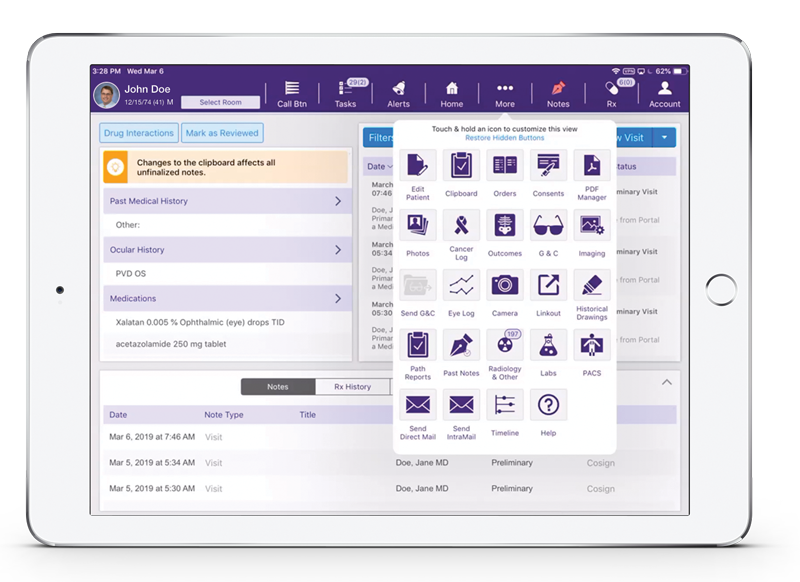
1. Accessible right from your ophthalmology EHR
Some ophthalmology image management systems require you to log in separately to use them, then search for the correct patient and date to find the right image. But when you’re in the exam room with a patient, you don’t want to have to follow any extra steps to view the image and make a diagnosis.
That’s why it’s essential to pick a system that interfaces with your ophthalmology EHR system, so the patient’s chart can actually link directly to the imaging study.
This means that with just one click, you can instantly pull up the images you need to support your diagnostic findings and clinical decision making. Then, you can easily switch back to documenting in your ophthalmic EMR without losing your place.
For instance, while you’re doing an interpretation, you can quickly switch to looking at your visual field reports. This level of convenience is hard to beat, and it’s something you’ll really miss if you don’t have it.
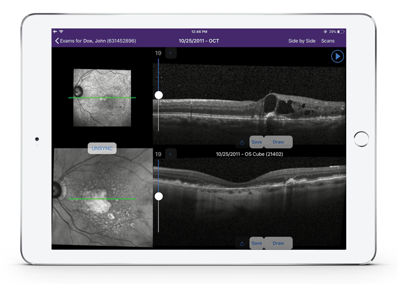
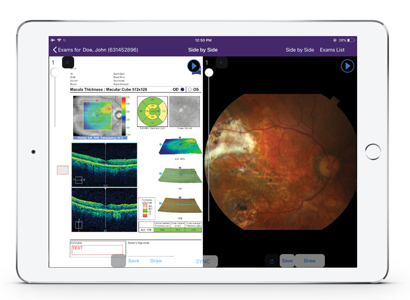
2. Serial comparison view with sync
A second feature you don’t want to live without is the ability to compare different images side by side.
Maybe you want to look at the right and left eyes, compare the same eye from two different dates, or see two different image types from the same date. Regardless of the situation, a comparison view can help give you stronger analytical capabilities.
When evaluating ophthalmic image management systems, you may find several that offer the basic ability to view images side by side. But when you look a little deeper, it may be harder to find some of the supporting capabilities that make the serial view much more useful and convenient.
I’ll give you an example: even if an image management system provides a side-by-side view, you may not find yourself keen to use it if you have to click through a great deal of options to get there. Instead, you want a system like Modernizing Medicine®’s Image Management, where you can enter comparison mode fast from the main view by simply selecting two images and clicking “compare.”
Also, when you’re viewing a single image, you want to be able to switch to a side-by-side view just as easily, without navigating back to the home view. It should take only a few clicks to find and select the second image.
Now, let’s say that you’re viewing a single image and you’ve zoomed in on a particular part of the eye. When you enter comparison mode from that single image view, the best image management software will automatically apply all the same options that you applied to the first image to both images.
So that means they’ll both be zoomed in on the bottom left of the eye. It’s a simple thing, but it’s incredible just how much faster and more convenient this is compared to adjusting all those settings manually.
And what happens if you want to view the top right of the eye from there? Well, if you leave sync mode on, the images will move together as you click and drag to view the top right. They’ll even zoom out together if you zoom out.
But if you’d like to move them separately—maybe you’re comparing different image types—you can simply turn off sync to adjust the settings individually.
As another handy option, you can click the arrows on the side of each image to move to the next image in the set. For instance, if you’re comparing a left eye fundus photo from last year to today, you can simply click the “next” arrows to move forward to the next image.
It’s all about convenience. With these powerful serial comparison view capabilities, you can save time and clicks when using diagnostic images to assess pathology and disease or treatment progression.
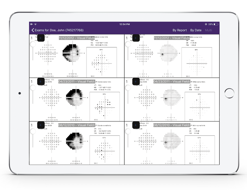
3. Visual field history comparison
Comparing two visual field images side by side is useful, but what about when you have three different dates to compare?
That’s where the visual field history feature comes in. This specialized view, available in Image Management for modmed® Ophthalmology, may be used to help you track progress of the right and left eyes together over three different visits.
When you use this viewer, the visual fields for right and left eye are displayed side by side with three different test dates showing one above the other. Since these images are specifically visual fields, they are automatically synced together, so they’ll all zoom and move together.
This allows you to see with a single glance what a specific part of the report looked like on each of your selected dates, saving you the time of having to pull up each report separately and scroll or zoom.
The dates you choose don’t have to be consecutive, either. So let’s say you’ve been seeing a patient for eight years. If you want to show them the gradual progression of their disease over time, you could compare the visual field report from eight years ago with the report from four years ago and from today. Or if you want to look at how it’s been progressing more recently, you could look at the report from two years ago, last year and today.
When it comes to analyzing historical visits, having access to this type of visual field history comparison view can be indispensable.
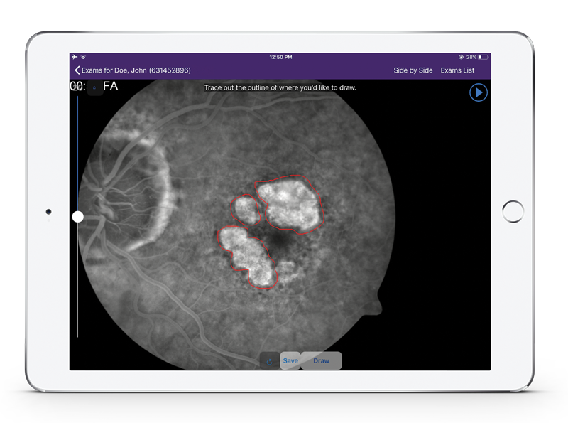
4. Easy ophthalmic image annotation
Number four on the list of vital ophthalmic image management features is being able to annotate your findings within the system, without having to go back to the diagnostic imaging device. There are three cases where this feature is particularly important:
- Supporting your diagnosis and billing codes
- Educating the patient about their condition
- Communicating with other healthcare professionals
Compared to just seeing you point at the screen (for patients) or reading your entire report (for healthcare professionals), looking at an annotation can be an easier, clearer way to understand your findings.
To give you more options and convenience when drawing, make sure to look for these capabilities as well:
- Select annotation color
- Draw freehand
- Place, rotate and resize arrows or circles
- Add text
- Undo previous actions
All these come standard with Modernizing Medicine’s ophthalmology Image Management software. Plus, annotated images are automatically saved as a separate file rather than overwriting the original file.
From there, you can add the annotated image directly into the patient’s record because Image Management is integrated with EMA for ophthalmology.
With these powerful annotation capabilities, it’s easy to illustrate the message you need to convey about the condition. And if patients have (literally) a clear picture of what’s going on with their condition, they just might be more likely to adhere to your prescribed treatment because they see how much it matters.
5. Seamless sharing inside and outside your practice
The features we’ve covered so far may sound great for you, the ophthalmologist, but what about other professionals at your practice and within the care continuum?
The truth is, if the right people can’t access the patient’s diagnostic images when they need them, it defeats the point of having them.
That’s why it’s essential to have an image management system that makes sharing easy.
Within your practice, this means a cloud-based system that provides centralized access across rooms, locations and team members. For instance, your ophthalmic tech can capture images in your imaging room, and you can find and review them while you’re in the room with the patient.
Or if you’re working at a satellite location, you don’t have to worry about being tethered to onsite servers or carrying physical copies of images with you. As long as you have a secure Internet connection, you’ll still have access to your images from the first location.
It should also be easy to share with healthcare professionals outside your practice, such as the patient’s primary care provider or a hospital system. With the touch of a button, Image Management lets you send image reports, including annotated images, as a fax or a digital PDF.
As stated earlier, sharing annotated images in addition to your written findings can help other healthcare professionals understand the images at a glance. In addition, you can share these annotated images with patients using your ophthalmology EHR’s patient portal to help with patient education.
With the ability to easily access and share images in an understandable format, you’re better able to communicate and help improve the continuum of care.
Conclusion
These features are just a few of many to consider when evaluating image management software, but they’re among the most important when it comes to your workflow.
If you can save even 5 clicks on a particular task, it will save you 250 clicks if you repeat that task 50 times a day. If you save 20 clicks per task, that number goes up to 1,000. Over time, this can add up to saving hours. Over the course of a year, it can add up to saving entire days.
By “doing your homework” and finding ophthalmology software that helps you move through your day faster, you can help free up more time to spend with your patients instead of your computer. And that’s a win-win for everyone.

Paul Gallogly, MD
Ophthalmology Team Lead, Posterior Segment
Dr. Paul Gallogly is the Ophthalmology Team Lead, Posterior Segment. He is a board-certified ophthalmologist who received his undergraduate and medical degrees from the University of Florida. He graduated with honors and was elected to the Alpha Omega Alpha Honor Medical Society. Dr. Gallogly completed an internship at Mount Sinai Medical Center in Miami Beach. Following his internship, he completed his residency in ophthalmology at Bascom Palmer Eye Institute in Miami.
In addition, Dr. Gallogly completed a fellowship in vitreoretinal diseases and surgery at the Bascom Palmer Eye Institute where he served as chief resident, Co-Director of Ocular Trauma and a member of the Residency Selection Committee. He is a member of the American Academy of Ophthalmology, Florida Society of Ophthalmology and the American Society of Retina Specialists. Dr. Gallogly has been an active participant in a number of clinical studies and has published articles in distinguished medical journals.


