


Ophthalmic Image Management
Access Your Diagnostic Images Virtually Anywhere, Anytime*
*with a secure network, meeting certain minimum requirements
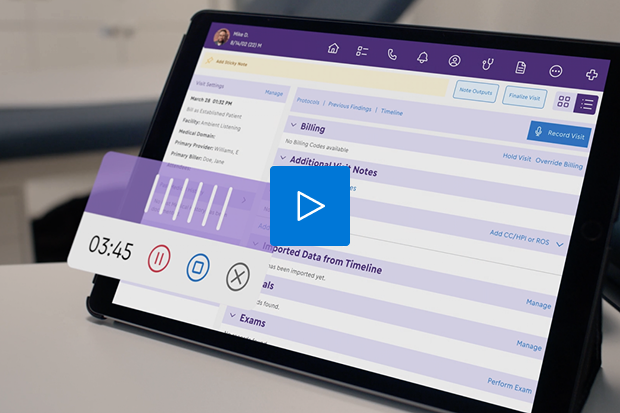
Whether you’re on the iPad or web, quickly open diagnostic images directly from the patient’s chart in our EMA® ophthalmology EHR. Image Management can automatically receive and store DICOM images from your connected ophthalmic diagnostic devices and provide centralized access to them in the cloud. When you can easily access, annotate, document and share images right from a current or previous encounter, you can work faster and improve care coordination.
Access Diagnostic Images With the Touch of a Button
Modernizing Medicine®’s Image Management system integrates with EMA, the #1 ophthalmology EHR¹, to help save you time and clicks. You can:
- Connect with advanced ophthalmology imaging devices
- Select images from exams you’ve just completed or from previous exams
- Sync and review image sequences side by side
With this flexible, modern system, the images to help support your diagnostic findings and documentation are exactly where you need them to be: within the patient’s chart.
¹2019 Black Book Market Research.
Why Image Management With EMA?
Our intuitive system brings the mobility and ease of use you love about EMA to your diagnostic image management.
Just like EMA, our Image Management system lets you use your iPad while talking to the patient and making eye contact. Plus, it makes a great tool for patient education, whether you’re performing initial diagnostics, treatment planning or a pre-surgery consultation.
Images are just a click away from your regular notes, so you can effortlessly switch back to documenting, then open the images again from within the patient’s chart if you need to. You can access patient images, document your findings and share the images with other healthcare professionals—all while the patient is still in the room.

Manage Images Right From the Patient’s Chart

Tired of spending more than a few seconds opening a patient image?
Within EMA, you can simply click once from the chart to view your diagnostic images organized by exam date and image type. From there, you can navigate intuitively, filter images, zoom in, annotate and more—all without losing your place.
You can jump from the patient’s chart to their images and back as fast as you can document the findings with EMA’s adaptive technology.
Review and Compare Images and Reports Side by Side
Our ophthalmology Image Management system makes it easy to view images right next to each other. You can:
- Compare historical data for the same exam type
- Review OCT scan data with intuitive slice-by-slice control
- View individualized scans with registration
- Switch from single image to comparison view without losing your filtered images, zoom or annotation tools
- Easily track progress over two or more visits with the visual field viewer comparison
- See OD and OS side by side automatically

Help Improve the Care Continuum

With our Image Management system, you can easily annotate images with freehand drawing, predefined shapes, text or redaction.
Since all the images are stored in the cloud, one specialist at your practice can capture a set of images, and another specialist can find them easily. Likewise, if you’re at a second location for one day a week, you’ll still have access to your images from the first location.